
Emerging Roles of Propolis: Antioxidant, Cardioprotective, and Antiangiogenic Actions
Abstract
Propolis has attracted attention in recent years due to its beneficial effects, which make it a potential preventive and therapeutic agent as well as a useful additive in food and cosmetics. The aim of this review is to discuss the growing evidence that propolis may, via a diverse array of biological actions, assist in the prevention of some inflammation-mediated pathologies including cardiovascular disease. The active components of propolis that have been identified so far include polyphenols and flavonoids. These compounds have cardioprotective, vasoprotective, antioxidant, antiatherosclerotic, anti-inflammatory and antiangiogenic actions. Many studies have been undertaken to elucidate the mechanism(s) by which propolis acts, which involve cellular signaling targets and interactions at the genomic level. This review will highlight the effects of propolis that may assist in the prevention of chronic degenerative diseases, such as cardiovascular disease.
1. Introduction
The growing market for natural products and alternative medicines has renewed interest in bee products, such as honey, royal jelly, pollen, and propolis [1, 2]. Propolis is the generic name for a complex resinous mixture collected by honey bees from the buds and exudates of various plants. Once collected, this material is enriched with saliva and enzyme-containing secretions and used in the construction, adaptation, and protection of hives [3, 4].
In recent years, many studies of the chemistry of propolis have been published, which reveal that its highly variable composition is influenced by the local flora at the collection site [5–7]. Although many biological activities of propolis are consistently observed, the components responsible vary between geographic and climatic zones [7].
There is considerable evidence on various chemical and biological aspects of propolis, but the therapeutic application and utilization by the pharmaceutical industry are still limited. This is mainly due to the variability of its chemical composition with geographical origin because bees utilize different plants in different ecosystems. Identification of the major compounds in propolis samples is essential; reports of the biological properties of propolis should include a detailed investigation of its composition and botanical sources [7, 8].
The constituents of propolis include polyphenols (flavonoids, phenolic acids, and esters), terpenoids, steroids, and amino acids [9]. There has been extensive research into the composition and biological activities of propolis from various countries [10–13]. Propolis samples from Europe, South America, and Asia have different compositions and therefore different biological activities [12, 14, 15]. However, propolis samples generally show great similarity in their overall composition, regardless of botanical source [15]. Brazilian red propolis has been found in two reports to contain high concentrations of phenolic compounds, 232 mg/g [16] and 257.98 mg/g, respectively [17]. Brazilian propolis also contained higher concentrations of total phenols than samples from other countries: China, mg/g [18] and mg/g [19]; Korea, mg/g [20]; Argentina, 187 mg/g [21]; India, mg/g [10]; Portugal, mg/g [22]; Cyprus, mg/g [23]; and Thailand, mg/g [19].
Studies indicate that propolis from Europe and China contains many flavonoids and phenolic acids; tropical propolis generally contains low concentrations of flavonoids [21, 24, 25].
Studies of propolis demonstrate the complexity of its composition and pharmacology; some compounds act independently, while others act synergistically. The therapeutic potential of propolis and its constituents has been the subject of many studies, which have established many pharmacological actions in preclinical testing.
In particular, propolis shows therapeutic potential and may have applications in the pharmaceutical and food processing industries [25–27]. Propolis reportedly has a range of biological activities, including immunomodulatory [28, 29], antibacterial [30], fungicidal [31, 32], anti-inflammatory, healing [33], analgesic/anesthetic [34, 35], and anticarcinogenic effects [36].
The relationship between oxidative stress, cardiovascular disease, and angiogenesis is well established. Events related to the pathophysiology of angiogenesis and associated cytokines and growth factors can lead to a poor prognosis in many diseases. In fact, chronic cardiovascular disease, oxidative stress, and angiogenesis are strongly associated with one another. In this review, we will present evidence that propolis extracts and their bioactive compounds have antioxidant, cardioprotective, and antiangiogenic activities (Figure 1).
2. Antioxidant Activity
It is well established that cellular metabolism generates reactive oxygen species (ROS), such as hydrogen peroxide (H 2 O 2 ), the superoxide anion ( ), and the highly reactive hydroxyl ion ( ), as well as reactive nitrogen species (RNS), especially nitric oxide (NO). ROS and RNS are ideal signaling molecules because they are locally generated, are highly and rapidly diffusible, and can be neutralized by cellular antioxidants [37, 38]. ROS are usually detoxified by intracellular enzymes, such as glutathione, superoxide dismutase, and catalase [39]. However, unbalanced production and degradation of ROS and RNS can result in accumulation of these reactive species, commonly referred to as oxidative stress. Exposure of macromolecules (lipid, proteins, DNA, etc.) to reactive species results in oxidative modifications with deleterious effects [40, 41].
The antioxidant capacity of propolis may be related to some of its biological effects, including chemoprevention. The flavonoids in propolis are powerful antioxidants, capable of scavenging free radicals and thereby protecting the cell membrane against lipid peroxidation [42]. Moreover, ROS and RNS, together with other factors, are involved in cellular ageing and death in conditions, such as cardiovascular disease, arthritis, cancer, diabetes, Parkinson’s disease, and Alzheimer’s disease [43–47]. Propolis can reduce cellular levels of H 2 O 2 and NO, which may be involved in its anti-inflammatory effects [48].
Diverse compounds from propolis have been described as potent inhibitors of oxidative stress. It is well known that the composition of propolis is variable; however, one of its major components, caffeic acid phenethyl ester (CAPE), blocks ROS production in several systems [49]. CAPE has also been identified as one of the major cancer chemopreventive and anti-inflammatory compounds in propolis. In vitro, propolis inhibits peroxidation of LDL and nitration of proteins. Moreover, in bovine aortic endothelial cells, propolis was reported to increase eNOS expression and inhibit NADPH oxidase (NOX) [50]. In vivo, propolis can increase antioxidant capacity in animals [51] and humans [52], leading to decreased lipid peroxidation, which is strongly associated with the risk of cardiovascular disease [53, 54]. Turkish propolis inhibited hydrogen peroxide (H 2 O 2 -) induced damage to DNA in cultured fibroblasts [55]. The antioxidant activity of phenolic components of the Turkish propolis may reduce damage to DNA induced by H 2 O 2 , which may be related to its chemopreventive activity. Red propolis from Cuba has shown protective effects in models of alcohol-induced liver damage, most likely due to its antioxidant properties [56]. Propolis inhibited macrophage apoptosis via effects on glutathione (GSH) and the tumor necrosis factors/nuclear factor kappa B (TNF/NF-κB) pathway [57, 58]. Moreover, Brazilian propolis from Baccharis dracunculifolia modulated 1,2-dimethlyhydrazine (DMH-) induced DNA damage in colon cells [59].
Isla et al. [60] described the protective effect of the Argentinian propolis from different sources against copper-mediated oxidative modification of lipids in unfractionated serum. Five types of Argentinian propolis, collected in different regions, inhibited lipid oxidation during the initiation and propagation phases. All five types of propolis diminished the maximal rate and extent of diene production, indicating that flavonoids can scavenge free radicals, such as superoxide [61, 62], protecting serum lipids from oxidation [60]. Jasprica et al. [52] showed that daily intake of powdered propolis for 15 days decreased the plasma malondialdehyde concentration in men. The extract (0.65 g), available in Croatian community pharmacies, contained 2.5% total flavonoids, equivalent to 16.25 mg of galangin. After 30 days of treatment, an increase in superoxide dismutase activity and changes in red blood cell parameters were detected, including cell count, hemoglobin and mean corpuscular volume, and cell distribution.
The antioxidant effect of Brazilian red propolis has been attributed to chalcones and isoflavonoids (including 7-O-methylvestitol, medicarpin, and 3,4,2′,3′-tetrahydrochalcone) that act as electron donors [63]. Furthermore, total flavonoid content in Brazilian red propolis is correlated with antioxidant activity, suggesting that all the phenolic and flavonoid compounds present contribute to this activity [64]. Chinese red propolis had a higher antioxidant activity than propolis from other sources, which was attributed predominantly to CAPE [65]. Chilean propolis also has antioxidant properties, which are correlated with its chemical composition [66]. Additionally, the antioxidant and free-radical-scavenging properties of propolis may be due to its phenylpropanoid content [67]. Thus, the available data indicate that propolis of different origins and distinct compositions consistently exhibit antioxidant actions. In addition to this antioxidant effect, bioactive compounds in propolis influence a large number of biochemical signaling pathways, and therefore physiological and pathological processes. Antioxidant capacity is one of the most important properties of propolis. Although there are several studies corroborating the potential antioxidant activity of propolis, there is no robust data on the safe dose in humans. Thus, there is need for clinical studies using propolis and its biologically active compounds, including studies of safety and bioavailability.
3. Cardioprotective Activity
The modulation of cardiovascular disease markers by propolis has been shown in several studies. In vitro and in vivo assays have been developed to elucidate the molecular mechanisms of this beneficial effect: regulation of glucose and lipoprotein metabolism; modulation of gene expression; decrease of the activity of scavenger receptors, inflammatory cytokines, and oxidative stress; improvement of endothelial function; and inhibition of platelet aggregation.
Atherosclerosis is a complex process involving the accumulation and modification of plasma lipoproteins in the arterial wall as well as the recruitment and proliferation of immune cells. This process advances through a series of stages beginning with the appearance of a fatty streak lesion, composed largely of foam cells, which are lipid-engulfed macrophages. The fatty streak evolves into a complex atherosclerotic plaque consisting of a lipid core covered by a fibrous cap, with some areas that are rich in inflammatory cells [68–70]. Several authors have postulated that dietary polyphenols reduce the risk of cardiovascular disorders and prevent the development of atheromatous plaques [71–73]. Thus, as a rich source of polyphenols, propolis represents a potential alternative strategy for the prevention of cardiovascular disorders.
Propolis has been shown to modulate lipid and lipoprotein metabolism. Propolis administration diminished liver cholesterol and triglyceride content and decreased the rate of hepatic triglyceride synthesis in rats [74, 75]. In LDL receptor knockout mice (LDLr−/−), treatment with Brazilian red propolis (250 mg/kg/day) decreased levels of triacylglycerol (TAG), total cholesterol (TC), and non-high-density lipoprotein cholesterol (non-HDL-C) [76]. LDLr−/− mice treated with Brazilian green propolis, which is rich in Artepillin C, pinocembrin, kaempferol, or with Chilean brown propolis, which is rich in pinocembrin, CAPE, quercetin, and galangin, also presented low levels of non-HDL-C. Moreover, mice treated with Brazilian red propolis showed significantly reduced TAG and TC, and increased HDL-C, compared to untreated mice. Furthermore, Turkish propolis, which is rich in flavonoids (mainly galangin, quercetin, kaempferol, apigenin, pinobanksin, pinocembrin, and pinostrobin) prevented alcohol-induced acute liver damage and lipid accumulation and induced beneficial changes in the serum lipid profile. HDL levels were high, and LDL levels were low, in mice treated with propolis and alcohol compared to alcohol only [59]. Moreover, propolis also positively affected HDL and LDL levels in rats. Treatment of diabetic rats with propolis of poplar origin diminished levels of total cholesterol, LDL-cholesterol, VLDL-cholesterol, and triglycerides, reinforcing the case that propolis modulates lipid metabolism and may be helpful in syndromes caused by blood lipid abnormalities [75].
In a recent study, the authors hypothesized that propolis may aid in the prevention rather than treatment of atherosclerosis. LDLr−/− mice were treated with distinct polyphenol-rich propolis extracts (250 mg polyphenols/mL/Kg) [76]. Brazilian green, Brazilian red, and Chilean brown propolis reduced the area of atherosclerotic lesions when administered preventively. The strongest inhibitory effect was observed for Brazilian red propolis, which also induced regression of atherosclerotic lesions [76]. Polyphenols from propolis inhibited the progression of atherosclerosis in LDLr−/− mice by improving the lipid profile and by downregulating proinflammatory cytokines, chemokines, and angiogenic factors. Propolis downregulated the mRNA expression of key genes involved in the atherosclerotic process, such as MCP-1, INFg, IL6, CD36, and TGFβ [76].
It is well known that the modification of the lipid profile is strongly associated with cardiovascular disease [76, 77]. Propolis diminished total cholesterol and elevated HDL-cholesterol in mice. One proposed mechanism of the hypocholesterolemic action of propolis involves the ABCA1 receptor. Many types of propolis upregulate ABCA1 gene expression, which is associated with increased HDL levels; thus, ABCA1 up-regulation may be one mechanism by which propolis improves the lipid profile [76].
An ethanolic extract of Brazilian red propolis (EERP) enhanced ABCA1 promoter activity in THP-1 macrophages [77]. Additionally, cholesterol efflux from macrophages to ApoA-I was significantly increased in a dose-dependent manner by EERP treatment. Thus, EERP significantly enhanced ApoA-I-mediated cholesterol efflux in THP-1 macrophages, which was accompanied by a marked up-regulation of the ABCA1 gene. The effect of EERP on ABCA1-dependent cholesterol efflux may be due to the activation of PPARγ and LXRα [77]. In HepG2 and Raw 264.7 cell lines, EEP promoted cholesterol efflux and increased the expression of ABCA1 and ABCG1. Accordingly, C57BL/6 mice treated with 50 mg/kg EEP once a day for 4 weeks by oral gavage showed increased plasma HDL-cholesterol but unchanged LDL-cholesterol [78]. Thus, in vitro and in vivo data suggest that the beneficial effects of propolis on lipid profile may be one of the mechanisms involved in its atheroprotective effects. This finding suggests that polyphenols from propolis may be useful for the prevention of atherosclerosis.
Platelet aggregation is a major contributor to the atherosclerotic process. Propolis components have shown important effects on platelet aggregation. CAPE (15 and 25 μM) markedly inhibited collagen-stimulated platelet aggregation. As CAPE is involved in various inhibitory pathways influencing platelet aggregation, it may be an important contributor to the potent antiplatelet actions of propolis [79].
NO is an important vasoactive mediator, with vasodilatory and antiaggregative actions that protect blood vessels when released from endothelial cells at low concentrations. However, when NO is produced in high concentrations by inflammatory cells, it may react with other nitrogen and oxygen species, inducing oxidative and/or nitrosative stress. Following the treatment of diabetic mice with poplar propolis, the levels of NO and nitric oxide synthase (NOS) decreased compared to nondiabetic mice [75]. Propolis decreases NO level by decreasing NOS activity, thus protecting blood vessel endothelial cells and reducing neuronal toxicity. Additionally, propolis exerts pharmacological effects by decreasing the actions of NO and PGE2 as well as by reducing the activation of protein kinase in diabetes [75, 80]. Moreover, ethanolic extracts of propolis (EEP) inhibit NO production by decreasing iNOS expression in Raw 264.7 macrophages and by directly inhibiting the catalytic activity of iNOS. The inhibitory effect of EEP on LPS plus IFN-g-induced NO production is mediated either by inhibition of iNOS gene transcription via an action on NF-κB sites in the iNOS promoter or by direct inhibition of the catalytic activity of iNOS [81]. As excess NO production has been implicated in the cardiovascular inflammatory process, the anti-inflammatory activities of EEP may also be mediated by modulation of NO levels.
It is well established that proliferation of vascular smooth muscle cells (VSMCs) is involved in the onset of atherosclerosis. Roos et al. [82] evaluated the antiproliferative activity of CAFE, one of the major components of propolis and honey-derived products, in primary rat aortic VSMCs stimulated by platelet-derived growth factor (PDGF). CAFE inhibited proliferation of VSMCs upon exposure to PDGF in a dose-dependent manner, by interfering with cell cycle progression from the G0/1- to the S-phase. This study indicates that the inhibition of smooth muscle cell proliferation may also be involved in the atheroprotective action of propolis.
4. Antiangiogenic Activity
Angiogenesis is the multistep process by which blood vessels are formed. This tightly regulated process involves the migration, proliferation, and differentiation of endothelial cells [83]. Regulation of angiogenesis is absent or aberrant in several diseases characterized by persistent, inappropriate blood vessel development. Inappropriate angiogenesis occurs in more than 80 diseases, particularly in many types of cancer and inflammatory diseases as atherosclerosis [84, 85].
According to Keshavarz et al. [86], green propolis extracts containing artepillin C and CAPE significantly reduced the number of new vessels formed and the expression of metalloproteinases (MMPs) and production of vascular endothelial growth factor (VEGF) from endothelial cells [87]. Different steps of angiogenesis can be affected by propolis and its components. Brazilian propolis and its major component, artepillin C, can inhibit proliferation of human umbilical vein endothelial cells (HUVEC), as well as endothelial cell migration and capillary tube formation, in a dose-dependent manner. Moreover, artepillin C can suppress angiogenesis in both in vivo and in vitro models, while CAPE inhibits MMP-2, MMP-9, and VEGF activity [87–90].
The effects of Brazilian propolis on HUVEC apoptosis were investigated by Xuan et al. [91]. At a low concentration (12.5 μg/mL), the polyphenols in ethanol extracts of Brazilian propolis decreased the expression of integrin b4 and p53 and the production of ROS. The opposite effects were observed at high polyphenol concentrations (25 and 50 μg/mL), along with depression of mitochondrial membrane potential. Thus, high doses of polyphenols from Brazilian propolis may induce HUVEC apoptosis by acting on the integrin b4 and p53 signaling pathway, resulting in disturbance of mitochondrial membrane potential and increased ROS generation.
Daleprane et al. [92] investigated the actions of polyphenols from Brazilian red propolis on models of angiogenesis. Brazilian red propolis is rich in 1,2,3-trimethoxy-5-(2-propenyl)-benzene, methoxyeugenol, homopterocarpin, medicarpin, 2,4,6-trimethylphenol, 49,7-dimethoxy-29-isoflavonol, 7,49-dihydroxyisoflavone, and 2H-1-benzopyran-7-ol [64]. Brazilian red propolis (10 mg/L) reduced the migration and sprouting of endothelial cells, attenuated the formation of new blood vessels, and decreased the differentiation of embryonic stem cells into CD31-positive cells. Moreover, Brazilian red propolis inhibited hypoxia- or dimethyloxalylglycine-induced mRNA and protein expression of the crucial angiogenesis promoter, vascular endothelial growth factor (VEGF), in a time-dependent manner [92].
Hypoxia is implicated in many inflammatory diseases. The proposal that hypoxia can induce inflammation has gained general acceptance from studies of the hypoxia signaling pathway [93]. Brazilian red propolis decreases accumulation of hypoxia-inducible factor 1 alpha (HIF1α) under hypoxic conditions, which in turn attenuates VEGF gene expression [92]. Reduced HIF1α protein half-life was associated with increased von Hippel-Lindau (pVHL-) dependent proteasomal degradation of HIF1α and reduced Cdc42 protein expression [92].
Brazilian green propolis extract, which is rich in artepillin C, was evaluated by Hattori et al. for its effects on cellular responses to hypoxia [94]. Five compounds that modulated HIF-1 activity were identified. Hydroxycinnamic acid derivatives from Brazilian green propolis inhibited not only HIF-1 transcriptional activity but also hypoxia-induced expression of HIF-1α protein and downstream target genes, such as Glucose transporter 1, Hexokinase 2, and vascular endothelial growth factor A. Furthermore, the HIF-1 inhibitors also inhibited angiogenesis. Daleprane et al. [76] investigated the effect of polyphenols from Brazilian propolis on angiogenic gene expression in atherosclerotic lesions of LDLr−/− mice, finding that angiopoietin I, angiopoietin II, VEGF, fibroblast growth factor, metalloproteinases 2 and 9, platelet-derived growth factor, and platelet endothelial cell adhesion molecule were downregulated by polyphenols from Brazilian red and green propolis.
It has been reported that the propolis extracts show antiproliferative activity and that both extracts induced cell death by necrosis [95]. The latter result indicates that certain compounds contained in propolis possess cytocidal activity based on necrosis rather than apoptosis. On the other hand, polyphenols, which are tumor necrosis factor-related, apoptosis inducing ligands, preferentially induce apoptosis in cancer cells and are not toxic to normal cells [96]. These results are therefore not consistent with each other. The inconsistency of propolis activity may be due to the presence of numerous compounds in varying levels, depending on their geographical origin. Generally, biological activity has been assessed by independent groups, making a direct comparison of their work difficult. Propolis-based medicines are often prepared from ethanol extracts of honey hive, as the extracts are generally water insoluble. Further studies are required to establish the quantity and safety control criteria for propolis to allow it to be used safely, and to gain the maximum benefit from its biological activities.
In vitro and in vivo studies are uncovering antiangiogenic activity in many natural health products, including propolis extracts and their constituents. Further preclinical research is required to determine whether individual compounds or complex mixtures will be optimal for clinical trials. A potential advantage of phytochemicals and other compounds from propolis is that they may act through multiple cellular signaling pathways, acting in different pathophysiological conditions, while also inhibiting angiogenesis and reducing inflammation. Overall, propolis constituents may be helpful as auxiliary therapies for diseases in which angiogenesis must be controlled, such as cancer and cardiovascular disease.
5. Perspectives on Propolis Utilization
Propolis contains a broad spectrum of compounds that have many biological activities. It is considered a useful product and is already used in alternative medicine. Recently, there has been a growing interest in its utilization by the food processing, cosmetic, and pharmaceutical industries. Considering this, further studies on the bioactive constituents of propolis are necessary to identify interactions mediating their biological effects. Further studies are also required on their bioavailability, stability in different preparations, and safe and effective doses for prevention or treatment of disease in animals and humans.
Conflict of Interests
The authors declare no conflict of interests.
Adhesive Behavior of Propolis on Different Substrates
Propolis is a sticky substance used by bees to seal their hive and protect the colony against pathogens. Its main components are plant resins, beeswax, essential oils, pollen, and other organic substances. The chemical and medicinal properties of propolis have been extensively studied, but little is known about its physical and especially adhesive properties. To gain a better understanding of propolis and its potential for adhesive applications, we performed several experiments, including adhesion tests with propolis in different conditions and on various substrates, differential scanning calorimetry analysis, and compression tests. Propolis shows clear viscoelastic behavior and temperature-dependent mechanical properties. Our results demonstrate that propolis adheres well to a wide range of substrates from glass to PTFE, but also enables stronger adhesion at higher temperatures and longer contact times. Even underwater, in wet conditions, quite a substantial adhesion was measured. The data are interpreted from a biomechanical point of view, and the significance of the obtained results for bee biology is discussed.
Introduction
Propolis is a sticky and ductile material that is produced by honeybees (Apis mellifera), mixing plant resins, wax, and other substances. Because of its strong adhesive nature, propolis is sometimes referred to as bee glue (Bankova et al., 2000; Bankova, 2005). Honeybees mainly forage resins from plant buds, but also have been reported to collect resin from tree barks and fruit surfaces (Alfonsus, 1933; Kumazawa et al., 2008; Simone-Finstrom and Spivak, 2010). Bees selectively choose what plants to acquire resin from (Isidorov et al., 2016). In Europe and North America poplars are believed to be the main source of resin for propolis (Greenaway et al., 1990; Bankova et al., 2000; Isidorov et al., 2016). Other plant sources for propolis production in temperate regions are aspen and birch (Isidorov et al., 2016). Since the contents of bud resins differ immensely between different plant species (Bankova, 2005), propolis contents and therefore properties are highly variable. In general, propolis consists of about 50% resin, 30% wax, 10% essential and aromatic oils, 5% pollen, and 5% of various other organic substances (Monti et al., 1983; Cirasino et al., 1987; Burdock, 1998). Meanwhile, more than 300 chemical components have been identified in propolis (Huang et al., 2014). The main constituents are phenolic compounds, such as flavonoids, aromatic acids and their esters (Bankova et al., 2000). Additionally, propolis contains phenolic aldehydes, ketones, terpenes, sugars, hydrocarbons, mineral elements, and enzymes (Bankova et al., 2000; Anjum et al., 2018).
Bees use propolis as a building material, for example to seal cracks and smooth out the internal walls of the hive (Burdock, 1998; Bankova et al., 2000). In addition to its mechanical functions, propolis has important chemical and bio-medical properties that protect the colony and contribute to social immunity (Simone-Finstrom and Spivak, 2010). Propolis possesses antibacterial, antifungal, antiviral, anti-inflammatory and hepatoprotective, antioxidant and antitumor properties (Anjum et al., 2018). Due to its pharmacological properties, propolis has been used by humans for multiple purposes (e.g., treating wounds or preserving corpses) throughout history (Ghisalberti, 1979; Anjum et al., 2018), and it still has manifold applications in medicine and cosmetics today (Burdock, 1998; Huang et al., 2014; Anjum et al., 2018).
Chemical and especially bio-medical properties of propolis have been studied extensively (Burdock, 1998; Cornara et al., 2017; Anjum et al., 2018), but little research has been done to understand its physico-chemical properties. A better understanding of the material properties of propolis could help biologists to understand how bees maintain such a sticky material. Beyond that, there is a strong potential for propolis applications in adhesive technology, as modern adhesive bonding technology is continuously searching for optimized environment-friendly adhesive solutions (Popov et al., 2017). Because of its pharmacological properties in addition to its stickiness, propolis might even be useful as a medical adhesive. Furthermore, knowledge about adhesive behavior of propolis on various surfaces could be used for developing anti-adhesive coatings.
The objective of this work was to characterize propolis mechanics, adhesion and other physico-chemical properties in order to ascertain whether the material might have other areas of application apart from its known medical uses.
Materials and Methods
Propolis Material
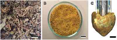
The raw propolis was provided by private beekeeper Dr. Oliver Schwarz (Stuttgart, Germany) (Figure 1A). Samples were harvested from beehives in autumn 2017 and spring 2018 by scraping the inside of hives, frames, and lids. Harvested propolis was stored outside in an unsealed container until summer 2018. To get consistent samples, the propolis chunks were frozen to −20°C, finely ground using a pre-cooled mortar and pestle, mixed, and subsequently stored at −20°C (Figure 1B). The pulverizing procedure was based on the method that was previously used to produce propolis extract (Bankova et al., 2016). To prevent contamination, propolis was only handled wearing gloves cleaned with ethanol (Rotipuran® ≥ 99.8%, p.a., Carl Roth GmbH & Co. KG, Karlsruhe, Germany).
FIGURE 1
Figure 1. Bee propolis. (A) Raw propolis as collected from the hive. (B) Homogenized propolis powder. (C) Cone-shaped propolis sample used for adhesion tests. Scale bar: 1 cm (A,B), 500 μm (C).
Density
Homogenized and kneaded propolis samples were weighed with a balance (AG204 DeltaRange®, Mettler-Toledo, Columbus, USA). The volume of the propolis sample was determined by measuring the volume of water it displaced in a 50 ml measuring cylinder. The samples density was then calculated by dividing the weight by the volume. Three samples were tested independently.
Differential Scanning Calorimetry
The thermal properties of propolis were studied using differential scanning calorimetry (DSC) (DSC 8500, Perkin Elmer, Waltham, USA). Aluminum pans and covers were used for sample preparation and closed manually bending the edges with tweezers. Raw propolis pieces of two harvests (Spring: 5.7 mg and Autumn: 6.8 mg) were analyzed separately. During the first heating cycle, propolis was heated from −50 to 60°C at 20 K/min. The temperature in the chamber was held for 1 min, then rapidly cooled down to −50°C at 200 K/min and held there for 4 min before continuing. For the second cycle, the sample was heated to 70°C at 20 K/min. Homogenized and kneaded propolis samples was also heated in a water bath, in order to observe melting behavior. Three samples were tested independently.
Compression Tests
Stress-strain curves were recorded during compression tests with propolis using a universal testing machine (Autograph AG-X plus, Shimadzu, Kyoto, Japan). Homogenized propolis was kneaded and subsequently filled into a flexible mold to be formed into propolis disks (6.5 mm high, 12.5 mm diameter). To ensure parallel and even surfaces on top and at the bottom of the disc, the discwas pressed between two smooth glass slides held apart by 6.5 mm high spacers on each side. In order to prevent the sample sticking to the compression plates, a piece of cling film was layered underneath and on top of the propolis disc. The sample was continuously loaded to a compression of 1% (engineering strain) and then unloaded. This process was repeated on the same sample for compressions of 5, 10, 20, and 30%, in order to see what degree of compression leads to elastic and which to plastic deformation. Measurements were carried out at different temperatures (4, 23, and 40°C), while applying and removing the load with a constant velocity of 4 mm/min, to examine the effect of temperature on elastic properties of propolis. The temperature was varied by cooling the sample and compression plate in the fridge, or heating it in an oven. Additional measurements were performed at different velocities (1, 2, and 4 mm/min) at a constant temperature of 23°C, in order to analyze the viscous behavior of propolis. Three samples were tested for each combination of temperatures and velocities. Stress-strain curves were generated to obtain information about the mechanical properties of propolis.
Weighing Experiments
Homogenized and manually formed propolis samples were weighed continuously over a period of 7 h using an ultra-microbalance (Sartorius® Cubis MSE2.7S, Sartorius AG, Göttingen, Germany). The lid of a reaction tube was cut off and weighed empty. For each measurement, there were three small propolis spheres, each weighing 40–50 mg, placed on the lid. These samples were weighed at intervals of 10 s for 7 h at 24°C and 45 % relative humidity (RH) (P330 temperature-humidity measuring instrument, Dostmann electronic GmbH, Wertheim-Reicholzheim, Germany). Three samples, each consisting of three propolis spheres, were weighed separately.
Adhesion of Propolis
Adhesion of propolis was tested on a clean, smooth glass surface. Just before each adhesion experiment, a small amount of homogenized propolis powder was defrosted and kneaded into a homogeneous mass. Cone-shaped propolis samples with a spherical tip were subsequently formed by hand wearing ethanol-cleaned gloves (Figure 1C and Supplementary Figure 2A). The topography of the sample was analyzed using a fast scanning 3D measurement microscope (Keyence VR 3100; Keyence Corporation, Osaka, Japan). The profile of the sample was measured in five positions arranged in a star shape through the highest point of the tip. To estimate the radius at the sample tip, a circle was fitted to the sample's profiles in five different orientations (Figure 2C). The circle's radii were measured and then averaged.
FIGURE 2
Figure 2. Adhesion experiments. (A) Experimental set-up for adhesion testing with Basalt-01 mechanical tester (Tetra GmbH). (B) Propolis contact in presence of fluid and without fluid. (C) 3D-profile of the propolis sample. The circle was used to measure the tip radius. The small subimage depicts the sample's topography, with darker areas depicted higher than lighter areas. (D) Typical force-distance curve obtained from adhesion experiments. FL, fluid droplet; FOS, fiber optical sensor; GC, glass capillary; MM, 2D-micro-manipulators; MR, mirror; MS, metal spring; PS, propolis sample; SU, substrate.
The effective elastic modulus and the pull-off force of propolis were measured with a microforce measurement device [Basalt-BT01; Tetra GmbH, Ilmenau, Germany (Gorb and Scherge, 2000; Jiao et al., 2000; Gorb et al., 2004)]. The device consists of micromanipulators as a platform holding the substrate material, a metal spring (springs with spring constants of 618 and 539 N/m were used) and a fiber-optical sensor (Figure 2A). The piezo-drive moves the sensor with the spring down for loading and up for unloading the sample. A shortened glass capillary (5 μl micropipette Blaubrand® Intra END, BRAND GMBH + CO KG, Wertheim, Germany) was attached to the metal spring with cyanoacrylate glue.
The freshly formed, cone-shaped sample of propolis was then mounted on the tip of the capillary without any additional glue. A glass slide (Standard microscopy slides (soda lime glass); Carl Roth GmbH + Co. KG, Karlsruhe, Germany) was cleaned with ethanol and distilled water. After drying out, the glass slide was fixed to the micromanipulator platform with double-sided adhesive tape to be used as substrate for subsequent adhesion tests (Article number 05338-00000-01, Tesa®, Norderstedt, Germany). The propolis sample was brought into contact with the substrate and retracted from the surface as soon as the load force reached 5 mN. The load was chosen to resemble the load applied by bees when handling propolis. As no studies exist on mandibular forces and pressures of honeybees, pressures previously measured at the tip of mandibles of predacious Coleoptera (Wheater and Evans, 1989) were used as a reference for the load applied to the propolis sample. Tip pressures were calculated as suggested by Wheater and Evans (1989):
P = F a A ( 1 )
where F a is the applied force and A is the contact area obtained from the contact radius.
Reference Measurements on Glass
On each propolis sample a set of 11 single measurements was performed, each on a different spot of the reference glass surface (N = 8 propolis samples, n = 11 measurements per sample). The last measurement of each set was carried out with a 60 s contact delay after loading and before unloading the sample to test the viscous properties of propolis. Experiments were carried out at room temperature (24.00 ± 0.53°C) and a relative humidity of 36.80 ± 9.01%. After the adhesion experiments, the substrate material was examined under a binocular microscope (Leica M205 A) in order to find possible propolis residues/prints in the contact area. Additional abbreviated reference measurements on glass (five repetitions) were performed with propolis samples used for tests on various other substrates described below.
Adhesion Under Different Tests Conditions
Some of the test conditions, described above for the reference measurements, were varied to prove whether they have an effect on propolis adhesion. First, repeated measurements were performed on the same spot. Second, measurements without prolonged contact time were performed to estimate the deformation of the sample tip after short contact measurements. Third, several measurements were carried out in fluid conditions, with a drop of distilled water (H 2 O) or oil (Mineral oil, light, Sigma-Aldrich, St. Louis, USA) being placed on the substrate (Figure 2B). Fourth, to study the influence of temperature on propolis adhesion, measurements were conducted at a higher room temperature, of 26°C. For each condition, 50–60 single measurements were performed (N = 5–6 propolis samples, n = 10 measurements per sample). Last, the experiments with an extended contact time of 60 s were performed to study the effect of contact time on adhesion. These experiments were performed in the last step of reference measurements on glass (N = 50 propolis samples, n = 1 measurement per sample).
Adhesion on Different Substrates
Various technical materials were used as substrates for subsequent adhesion experiments. A polytetrafluoroethylene (PTFE) plate (Technische Materialien Katarzyna Kowalewska, Görlitz, Germany) and a steel plate (EN 1.4016, Abrams Premium Stahl®, Osnabrück, Germany) were polished using a polishing machine (Minitech 233; Presi, Eybens, France) with alumina oxide suspensions using polishing papers with descending particle sizes (12, 3, 1, and 0.3 μm) to achieve similar and minimal surface roughness for all substrates (Supplementary Figure 1). They were cleaned with ethanol and distilled water and dried prior to usage in the experiments. A resin replica of a smooth, clean glass surface was prepared by a two-step molding method (Gorb, 2007; Koch et al., 2008). The negative template of the glass slide was produced using a two-component dental wax (Affinis light body, ISO 4823, polyvinylsiloxane, Coltène Whaledent AG, Altstätten, Switzerland) and filled with resin (Spurr's low viscosity kit, Plano, Wetzlar, Germany) that was subsequently polymerized at 70°C for 48 h. This resin substrate will be further referred to as ”Spurr.”
Tests on these technical substrate materials were performed as described for the reference glass substrate. After 10 measurements on the substrate material, five additional reference measurements were carried out on glass. For each technical substrate material, 50–60 single measurements on different sites were performed (N = 5–6 propolis samples, n = 10 measurements on substrate per sample).
Examination of Surfaces
The contact angles of water, diiodomethane, and ethylene glycol on the reference glass surface, as well as of the PTFE, steel and Spurr substrates were measured according to the sessile drop method (2 μl drop volume) using an optical contact angle measuring system (OCAH200, DataPhysics Instruments GmbH, Filderstadt, Germany). For each substrate, 5–10 contact angle measurements were conducted for each liquid. The substrate's surface free energy and its dispersive and polar components were calculated according to the method by Owens and Wendt (1969).
The substrate's surface roughness was measured by a confocal 3D laser scanning microscope (Keyence VK-X250; Keyence Corporation, Osaka, Japan). The corresponding Multi File Analyzer software (Version 1.2.6.106, Keyence Corporation, Osaka, Japan) was used to obtain the following roughness parameters: arithmetical mean height of the surface (S a ), maximum height of the surface (S z ), texture aspect ratio of the surface (S tr ), arithmetic mean peak curvature (S pc ), and developed area ratio (S dr ).
Additionally, for surface examination, standard light microscopy (Leica M205 A, Leica Microsystems Inc., Wetzlar, Germany) and 3D surface profilometry (Keyence VR 3100, Neu-Isenburg, Germany) were used.
Data Analysis
Adhesion experiments were evaluated using Matlab (version R2015b, The MathWorks, Inc., Natick, USA). The unloading part of force-distance curves (Figure 2D) acquired from adhesion experiments was fitted according to the JKR theory (Johnson et al., 1971) (Supplementary Figure 2B).
a 3 = 3 R 4 E [ F a + 3 π R γ + 6 π R F a γ + ( 3 π R γ ) 2 ] ( 2 )
where a is the contact radius, F a is the applied load, R is the tip radius, E and Δγ are the effective elastic modulus and the work of adhesion, respectively.
The work of adhesion Δγ is the energy per unit of area needed to separate two bodies in contact. It was chosen as a measure of adhesion, because it is independent of the contact area. Work of adhesion (Δγ) was estimated from the unloading curve:
Δ γ = - 2 F 3 π R ( 3 )
where F is the pull-off force and R is the tip radius.
To characterize viscoelastic properties of propolis, a generalized Maxwell model was used (Christensen, 1982). The sample's viscosity was estimated from experimental force curves using the following equation (Cheng et al., 2005; Kovalev et al., 2018):
F a = 4 R d 1 . 5 3 ( 1 - ν 2 ) ( E ∞ + E 1 e - E 1 t η 1 + E 2 e - E 2 t η 2 ) ( 4 )
where d is the displacement, t is the time under load, E ∞ /E 1 /E 2 and η 1 /η 2 are the Young's moduli and viscosities of the static and two dynamic components, correspondingly ν is the Poisson ratio assumed to be equal to 0.49 (Kovalev et al., 2018).
Statistics
The data were statistically analyzed using the software R, version 3.6.1 (The R Foundation for Statistical Computing, 2019). Data was tested for normal distribution and variance homogeneity using Kolmogorov-Smirnov and Levene's tests, respectively. The comparison of propolis adhesion under different conditions and on different substrates was performed with a one-way ANOVA and a pairwise multiple comparison procedure (Tukey test). An unpaired two-sample t-test was performed to compare the mean Young's modulus of propolis at 24 and 26°C. Correlation analysis of Young's modulus and work of adhesion obtained from adhesion experiments was performed by calculating the Pearson's correlation coefficient.
Results
Density of Propolis
Homogenized and kneaded pieces of propolis were weighed. Subsequently, the volume of weighed pieces was determined by measuring the volume of water it displaced. The propolis sample did not dissolve or absorb water during the experiment. The density of propolis was calculated to be 0.953 ± 0.001 g/cm3 (N = 3).
Thermal Behavior of Propolis
Melting of Propolis
When heating propolis in a water bath to 60–70°C, its separation into two phases was observed (N = 3). A phase resembling beeswax melted and turned into a transparent fluid with a yellow tint, while the other phase remained more viscous and dark brown. After once being heated to between 50 and 60°C, cooled down propolis components turned hard at room temperature and did not return to their original malleable state.
Differential Scanning Calorimetry
A DSC analysis of raw, unhomogenized propolis harvested in spring and autumn was performed to further investigate thermal properties of propolis. During the heating cycles, the variation of the heat flow as a function of the temperature revealed endothermic phase transitions at 54–55°C and 64°C (Figure 3A). No distinct differences between the samples from different harvests were found.
FIGURE 3
Figure 3. Thermal properties of propolis. (A) DSC analysis of propolis. (B) Stress-strain curves obtained from compression tests with propolis at different temperatures.
Compression Tests at Different Temperatures
The hardness of propolis changed depending on temperature. It is hard and brittle at temperatures below 10°C, allowing it to be broken or ground to a fine powder. At room temperature, propolisis is malleable and tacky. When the temperature rises above room temperature, propolis becomes increasingly softer and tackier. These changes were reflected in stress-strain curves obtained from compression tests (Figure 3B, N = 3). Compared to tests performed at 23°C, ~4 times higher stresses were necessary at 4°C to achieve the same strain. Furthermore, at 40°C for the same strain the stresses were roughly 10 times lower than that at 23°C. In compression tests, performed at 20–800 kPa stress and temperature 4–40°C, visco-plastic deformation of propolis was observed.
Weighing Experiment
A hardened outer layer formed on propolis samples stored at room temperature for several hours, though the inside of the propolis samples remained softer. To characterize the process of volatile components evaporations, the weight of the propolis samples was measured over time. On average, the samples lost 0.9 ± 0.3% (N = 3) in mass over a period of seven hours at room temperature of 24°C (Figure 4).
FIGURE 4
Figure 4. Display of weighing experiments performed on propolis. The curve shows the mean weight of the samples (N = 3) over time. The gray area represents standard deviations at corresponding time points of measurement.
Adhesion of Propolis
Cone-shaped propolis samples that were used for adhesion experiments had a mean tip radius of 182.08 ± 55.18 μm (N = 135) (Figure 2C). The samples were brought into contact with different substrates at a mean applied normal force of 5.2 ± 0.7 mN. The contact radius at maximum load was calculated to be 36.06 ± 17.14 μm, using Equation (3). Mean pressure at the propolis sample was calculated to be 1.85 MPa. At a room temperature of 24°C, propolis samples had a mean Young's modulus of 11.23 ± 6.77 MPa (N = 45). Compared to measurements at 24°C, a 2°C increase in temperature resulted in a significantly lower elastic modulus of 6.55 ± 4.89 MPa (N = 10, P = 0.044). In adhesion experiments, performed with propolis on a glass surface, the mean pull-off force was 2.12 ± 0.77 mN and the mean work of adhesion was calculated to be 2.96 ± 1.27 J/m2 using Equation (3). According to Pearson's correlation test, Young's modulus and work of adhesion of propolis are anti-correlated (r = −0.999, p < 0.01). The mode of failure during experiments was adhesive, since examination of the substrate surfaces after adhesion experiments using light microscope showed no propolis residues in the contact area. 3D surface profilometry of the sample's tip before and after the experiment also showed no shape change and therefore no plastic deformation occurred during testing at short contact times. However, at a contact time of 60 s, samples clearly exhibited viscoplastic deformation, since the tip area was considerably flattened.
Propolis Adhesion Under Different Conditions
Propolis adhesion was subsequently measured on a glass surface under different conditions and compared by performing a one-way ANOVA (Figure 5A, Table 1). The P-value was found to be smaller than the significance level of 0.01. Therefore, a post-hoc Tukey test was conducted to find pairwise differences. P-values smaller than 0.05 were considered significant. The work of adhesion of propolis did not depend (1) on the contact area and (2) on whether the measurements were repeatedly done at the same location or each measurement was done at new location (3.08 ± 0.79 J/m2, P = 0.9998).
FIGURE 5
Figure 5. Propolis adhesion. (A) Adhesion of propolis on glass under various conditions. (B) Adhesion of propolis on different substrates. Experiments were conducted using Basalt-1 mechanical tester (Tetra GmbH, Ilmenau, Germany). If not stated differently, tests were carried out at room temperature (24°C) with a set loading force of 5 mN and with every individual measurement performed on a different location on the substrate (N = 5–6 propolis samples per condition or substrate, n = 10 individual measurements per sample). Box plots show the median value (line), the ends of the boxes define the 25 and 75th percentiles, and the error bars the 10 and 90th percentiles. The outliers are shown as black dots. Conditions and substrates marked with different letters differ significantly from each other (one-way ANOVA, P < 0.001 and Tukey test, P < 0.05).
TABLE 1
Table 1. Propolis adhesion in different conditions.
Propolis adhesion in oil and water was measured. Propolis adhered to glass, even underwater and in oil, however, the work of adhesion measured (0.86 ± 0.47 J/m2) was significantly lower than that in the dry condition (P < 0.0001). No significant difference between the work of adhesion in water and in oil was found (P= 1.0). Raising the temperature from 24 to 26°C, the work of adhesion of propolis increased significantly to 4.67 ± 1.36 J/m2 (P < 0.0001). Some experiments performed at 26°C were abandoned due to cohesive failure and plastic deformation of the propolis sample after a few single measurements. Significantly higher work of adhesion of 6.72 ± 1.75 J/m2 also occurred, when increasing the contact time at maximum load to 60 s (P < 0.0001).
Viscoelastic Behavior
As stated above, elongated contact times led to plastic deformation of the propolis samples and to a decrease of the load force overtime due to material relaxation (N = 50). The force-time curve was fitted with the exponential function (Bankova et al., 2000) with either one or two exponents in the 60 s contact regime (Figure 6A). Table 2 shows the fit parameters for one representative force-time curve. For all measurements a better fit was achieved with two exponents. In compression tests, propolis behaved differently depending on the velocity the load was applied with. At higher velocities, higher stresses had to be applied to achieve the same strain (Figure 6B). No elastic deformation occurred in compression tests performed using a universal testing machine.
FIGURE 6
Figure 6. Viscoelasticity of propolis. (A) Typical force-time curve obtained from adhesion tests with propolis on glass with a 60 s delay between loading and unloading. (B) Stress-strain curves obtained from compression tests with propolis at different velocities (1, 2, and 4 mm/min).
TABLE 2
Table 2. Fit parameters to estimate the viscoelasticity of a representative propolis sample using the Equation (4).
Adhesion to Different Substrates
To test the effect of substrate materials on propolis adhesion, adhesion tests were performed with several substrates: PTFE, steel, and Spurr's resin with different surface free energies. All tested substrates were smooth with an arithmetical mean height of the surface (Sa) between 0.034 and 0.042 μm (Supplementary Table 1), though their surface energies differed (Supplementary Table 2). PTFE had the lowest surface energy of 16.7 mJ/m2, then Spurr with 28.08 mJ/m2, and steel with 37.92 mJ/m2, while glass had the highest surface energy of 58.25 mJ/m2 among the tested materials. Despite the different surface energies, the work of adhesion obtained from adhesion experiments with propolis was similar for all tested substrate materials and ranged between 2.29 and 3.61 J/m2 (Figure 5B, Table 3). The highest work of adhesion was measured for Spurr epoxy resin, the lowest on steel. Statistically significant differences were only revealed for work of adhesion between glass and Spurr (P = 0.0049), as well as between steel and Spurr (P = 0.0004).
TABLE 3
Table 3. Propolis adhesion on different substrates.
Discussion
Chemical composition and medicinal use of propolis have previously been studied (Burdock, 1998; Anjum et al., 2018), but no in-depth analysis of adhesive and other physical material properties of propolis have been conducted so far. To be able to better understand how bees handle this sticky material, propolis was characterized as a biological adhesive in this paper. Accordingly, thermal properties, viscosity, elastic modulus and density mattered as important parameters in this study in addition to propolis adhesion itself (Kellar, 2011). From the density measurements, we can conclude that the density of propolis (0.953 g/cm3) is very close to the density of beeswax 0.957 g/cm3 reported in (Bernal et al., 2005). Such a density match might simplify the manufacture and handling of propolis by honeybees.
Thermal Properties of Propolis
Propolis has previously been described to melt at temperatures between 60 and 70°C (Krell, 1996; Wagh, 2013). When heated in a water bath in our experiments, the beeswax component of propolis melted at 60–70°C. The resin component of propolis only softened, but did not become fluid. In our DSC analysis, propolis demonstrated a phase transition at 63°C. This corresponds to the melting temperature of beeswax at 62–66°C depending on its origin (Morgan et al., 2002; Gaillard et al., 2011). During the heating process, another phase transition occurred at about 55°C. Three polymorphic transitions during the heating of beeswax have previously been identified, one corresponding to the peak at 55°C (Gaillard et al., 2011). The other two transitions were not visible in our DSC results, probably due to a higher heating speed of 20 K/min compared to 1 K/min used by Gaillard et al. (2011). Previous DSC studies performed on beeswax/rosin mixtures also showed that a higher resin content leads to a decrease in the total heat flow and less pronounced or lacking secondary peaks corresponding to phase transitions (Gaillard et al., 2011). Amorphous resins, like rosin gum, often do not exhibit a clear melting point, but soften over a wide temperature range until they become liquid (Gaillard et al., 2011), while volatile essential oils usually have low melting points (−0.79°C for essential oil of cassia) (Ghodki and Goswami, 2016). This could explain why no additional peaks are present in the DSC thermograms apart from those corresponding to the beeswax component of propolis. Propolis has been reported to be hard and brittle when cold (under 15°C) (Krell, 1996; Wagh, 2013) and increasingly soft and sticky, when heated above 45°C (Krell, 1996; Wagh, 2013). This was confirmed by our adhesion and compression tests. Propolis behaved distinctly different in compression tests conducted at 0, 23, and 40°C and with 67 μm/s compression speed. Compared to tests performed at lower temperatures, a considerably lower force was needed at 40°C, to achieve the same level of compression. Both propolis elasticity modulus and viscosity decrease with an increasing temperature. Compression tests with beeswax at 3, 24, and 34°C revealed similar behavior (Morgan et al., 2002).
Young's Modulus of Propolis
At 24°C, propolis has an elastic modulus of 11.2 MPa, which resembles elastic modulus of rubber (Smith, 2016). The Young's modulus, however, was very variable between samples, although all were formed from the same batch of homogenized propolis. Presumably, small changes in temperature, humidity, or/and kneading procedure had an influence on the modulus. Additionally, local inhomogeneities within the material might influence the modulus, since homogenized propolis consists of many heterogeneous microparticles, the size of which is comparable to the size of the tip (contact area). A small increase in temperature of 2°C decreased the Young's modulus of propolis significantly from 11.2 to 6.6 MPa. This also supports the observation made during compression tests at 40°C and when handling propolis that propolis becomes softer at higher temperatures. The elastic modulus of beeswax at room temperature was reported to be 39 MPa (Hossain et al., 2009). The addition of plant bud resins, oils, and other components, thus results in a softer and more pliable material compared to the pure beeswax (at room temperature).
Viscoelastic Behavior of Propolis
Viscoelastic materials show combined elastic and viscous responses under mechanical stress (Ferry, 1980). We found that propolis exhibited viscoelastic behavior. Deformation and relaxation of viscoelastic materials is time-dependent (Kellar, 2011). This became clear, as propolis deformed elastically during adhesion experiments with short contact times, but viscoelastically, when contact times were extended to 60 s. Elastic contact could be distinguished from plastic contact, as the distance of the sample tip to the substrate and the tip profile did not change between single measurements. The multiple components of propolis also affect its viscoelastic behavior. During extended contact time, the measured loading force gradually decreased over time as the material relaxed. The relaxation curve of propolis was best fitted with an exponential function with two exponents, which is indicative of a composite material with at least two components (Xie et al., 2011). This result is not surprising, since propolis mainly composed of resin (50%) and wax (30%) (Burdock, 1998).
Compression tests at different velocities showed that the material behaved stiffer at higher velocities and more force had to be applied in order to deform it. This rate dependence due to non-Newtonian properties is typical for viscoelastic materials (Smith, 2016).
Hardening of Propolis
After heating propolis over 50–60°C and cooling down, it hardened considerably, and when exposed to the room temperature a hardened layer formed on the outside of the sample over the course of a day. It has previously been reported that propolis hardens over time, becoming brittle and darker in color (Teslenko et al., 2014). A possible reason for hardening could be the evaporation of water or/and volatile components (Kellar, 2011), as weighing propolis over 7 h showed that samples lost ~0.9% in weight over that period of time. Volatile propolis components, such as mono- and sesquiterpenoids, represent about 10% of propolis constituents and their loss could be responsible for propolis hardening (Bankova et al., 2000; Huang et al., 2014). Some resins also harden over time due to polymerization of diterpenoids initiated by light and oxidation (Langenheim, 1990). While beeswax has not been reported to harden, it is therefore likely that the resin component is responsible for the hardening of propolis.
Adhesion of Propolis
The work of adhesion calculated from the pull-off force was chosen as the measure for propolis adhesion, as it provides comparable values that do not depend on the sample radius. This made it possible to compare the results of adhesion tests conducted with propolis at different conditions and on various substrates. Propolis adhesion on glass served as a reference and was measured to be ~3 J/m2. In a previous study, work of adhesion of 3.56 J/m2 was reported for a blend of 75% rosin and 25% beeswax (Girard et al., 2014). This material resembles propolis, as it is also a mixture of beeswax and resinous substances in a comparable ratio, and it exhibits similar work of adhesion. The mode of failure of propolis during adhesion experiments was rather adhesive than cohesive, as no visible propolis residues were found in the contact area with the substrate.
Adhesion at Various Conditions
Experiments that were conducted repeatedly on the same spot of the substrate further confirmed the assumption that adhesive failure occurs. There was no difference in adhesion between reference measurements and measurements on the same spot, as could be expected if cohesive failure occurs and material residues accumulate in the contact area and tip geometry changes.
Furthermore, propolis also adhered to a glass surface in presence of fluid (oil or water), although here adhesion was significantly lower than that in dry conditions. Most industrial adhesives are not able to adhere in flooded conditions (von Byern and Grunwald, 2010). However, biocompatible glues able to adhere in wet conditions are needed for many biomedical applications (von Byern and Grunwald, 2010). The ability of propolis to adhere under water could therefore be an interesting property worth investigating further.
Significantly higher propolis adhesion was observed when the temperature was increased by only 2°C from to 24 to 26°C. It has been previously described that propolis becomes increasingly soft and sticky, when heated above 45°C (Krell, 1996; Wagh, 2013), but it seems that this process starts at even lower temperatures. At higher temperatures, the Young's modulus of propolis decreased and the material became softer. Adhesion energy is theoretically independent on Youngs' modulus. However, an elastically soft material can adapt to the substrate surface and thereby increase the real contact area and adhesion (Lorenz et al., 2013). Further experiments are necessary to study propolis adhesion at even higher temperatures and further extend our knowledge about this material, since bees handle propolis in the hive at temperatures usually ranging between 32 and 36°C (Jones, 2004). Higher adhesion of propolis was also measured in experiments with longer contact times, because this viscoelastic material had time to flow and adapt to the substrate surface profile, increasing therefore both the contact area and work of adhesion. This effect of time-dependent deformations has previously been reported (Reitsma et al., 2000). The viscoelastic deformation was also visible in the profile of propolis samples after experiments with prolonged contact times.
Adhesion on Various Substrates
Substrate surface energy usually has a strong effect on adhesion (Gorb and Heepe, 2017). In our adhesion experiments, conducted on various smooth substrates with different surface energies [glass: 64 mJ/m2 (Weng et al., 2016), PTFE: 19 mJ/m2 (Kinloch, 1987), steel: 40 mJ/m2 (Mantel and Wightman, 1994)], the differences in adhesion forces were lower as expected. Substrates with low surface energy, such as PTFE, have low Hamaker constant and polarizability, and affect the strength of van der Waals interactions, which is why lower adhesion would usually be expected on these surfaces (Gorb and Heepe, 2017). However, propolis adhered more or less similarly to all tested substrates. There was no significant difference between adhesion on a polar glass surface and an unpolar PTFE substrate. Low dependence on substrate chemistry is often a characteristic of biological adhesives (Richter et al., 2018). We assume that some fluid components (e.g., mono- and sesquiterpenoids) of propolis may alter the interface between propolis sample and substrate and make adhesion almost independent of the substrate. These fluids might also be responsible for enlarged real contact area. Chemical characteristics of propolis should be further investigated to better understand their influence on propolis adhesion.
General Conclusions and Outlook
Exact composition of propolis can vary immensely depending on environmental conditions, resin sources, bee colony, season, etc. (Bankova, 2005; Mello and Hubinger, 2012). One can expect that the variability of propolis also extends to its physical and adhesive properties. The presented results are therefore a starting point for further experiments with different propolis types, as well as on propolis components, such as bud resins and different bee waxes.
The results of this work show that the composite nature of propolis is reflected in many of its physical properties, such as viscoelasticity, Young's modulus, and adhesion. Different components of propolis, mainly plant resins and beeswax, have long been used in various adhesives due to their characteristic properties. Resin is a good adhesive that can be added to adhesive mixtures, to provide tack and peel (Richter et al., 2018). In contrast, beeswax does not have initial tack, but it can act as a softener for adhesive formulations (Richter et al., 2018). Combinations of a resin (rosin) and beeswax have been used as adhesives since the Mesolithic period and have also previously been studied for their thermal and mechanical properties (Gaillard et al., 2011; Girard et al., 2014).
As propolis was found to adhere to a wide range of substrates and even in underwater and flooded oil conditions, it might be interesting for numerous adhesive applications in the future. Recently, the interest in bio-inspired adhesives has strongly increased due to the growing need for sustainable or environment-friendly adhesive solutions (Richter et al., 2018). For example, adhesives used for medical applications often have to serve different purposes, such as high tack, moisture resistance, durability, biocompatibility, and removability (von Byern and Grunwald, 2010), which all could at least partly be fulfilled by propolis. The antimicrobial effect of propolis might also be an advantageous additional property for future medical adhesives.
Data Availability Statement
The raw data supporting the conclusions of this article will be made available by the authors, without undue reservation.
Author Contributions
LS, JS, OS, SG, and AK contributed to conception and design of the study. LS conducted and evaluated the experiments and wrote the first draft of the manuscript. KW performed and evaluated the DSC analysis. All authors contributed to manuscript revision, read, and approved the submitted version.
Conflict of Interest
The authors declare that the research was conducted in the absence of any commercial or financial relationships that could be construed as a potential conflict of interest.
Supplementary Material
The Supplementary Material for this article can be found online at:
References
Alfonsus, E. C. (1933). Some sources of propolis. Glean Bee Cult. 61, 92–93.
Bankova, V., Bertelli, D., Borba, R., Conti, B. J., da Silva Cunha, I. B., Danert, C., et al. (2016). Standard methods for Apis mellifera propolis research. J. Apic Res. 58, 1–49. doi: 10.1080/00218839.2016.1222661 CrossRef Full Text | Google Scholar
Bankova, V., de Castro, S., and Marcucci, M. (2000). Propolis: recent advances in chemistry and plant origin. Apidologie 31, 3–15. doi: 10.1051/apido:2000102 CrossRef Full Text | Google Scholar
Bernal, J. L., Jiménez, J. J., del Nozal, M. J., Toribio, L., and Martin, M. T. (2005). Physico-chemical parameters for the characterization of pure beeswax and detection of adulterations. Eur. J. Lipid Sci. Technol. 107, 158–166. doi: 10.1002/ejlt.200401105 CrossRef Full Text | Google Scholar
Christensen, R. (1982). Theory of Viscoelasticity, 2nd Edn. London: Academic Press; doi: 10.1016/B978-0-12-174252-2.50012-0 CrossRef Full Text
Ferry, J. D. (1980). Viscoelastic Properties of Polymers. New York, NY: Wiley-Blackwell. Google Scholar
Gaillard, Y., Mija, A., Burr, A., Darque-Ceretti, E., Felder, E., and Sbirrazzuoli, N. (2011). Green material composites from renewable resources: polymorphic transitions and phase diagram of beeswax rosin resin. Thermochim. Acta 521, 90–97. doi: CrossRef Full Text | Google Scholar
Ghodki, B. M., and Goswami, T. K. (2016). Physico-thermal and flavoring characteristics of essential oil of cassia. J. Essent. Oil Bear Plants 19, 854–862. doi: 10.1080/0972060X.2016.1187091 CrossRef Full Text | Google Scholar
Gorb, E., Kastner, V., Peressadko, A., Arzt, E., Gaume, L., Rowe, N., et al. (2004). Structure and properties of the glandular surface in the digestive zone of the pitcher in the carnivorous plant Nepenthes ventrata and its role in insect trapping and retention. J. Exp. Biol. 207, 2947–2963. doi: 10.1242/jeb.01128 PubMed Abstract | CrossRef Full Text | Google Scholar
Gorb, S. N., and Heepe, L. (2017). “Biological fibrillar adhesives: functional principles and biomimetic applications,” in Handbook of Adhesion Technology, eds L. F. M. da Silva, A. Ochsner, and R. D. Adam (Cham: Springer International Publishing), 1–37. doi: 10.1007/978-3-319-42087-5_54-2 CrossRef Full Text | Google Scholar
Gorb, S. N., and Scherge, M. (2000). Biological microtribology: anisotropy in frictional forces of orthopteran attachment pads reflects the ultrastructure of a highly deformable material. Proc. R Soc. London Ser. B Biol. Sci. 267, 1239–1244. doi: 10.1098/rspb.2000.1133 PubMed Abstract | CrossRef Full Text | Google Scholar
Greenaway, W., Scaysbrook, T., and Whatley, F. R. (1990). The composition and plant origins of propolis: a report of work at Oxford. Bee World 71, 107–118. doi: 10.1080/0005772X.1990.11099047 CrossRef Full Text | Google Scholar
Hossain, M., Ketata, C., and Islam, M. R. (2009). “Experimental study of physical and mechanical properties of natural and synthetic waxes using uniaxial compressive strength test,” in Proceeding of Third International Conference on Modeling, Simulations and Applied Optimization, Sharjah, United Arab Emirates, 1–5.
Johnson, K. L., Kendall, K., and Roberts, A. D. (1971). Surface energy and the contact of elastic solids. Proc. R Soc. A Math. Phys. Eng. Sci. 324, 301–313. doi: 10.1098/rspa.1971.0141 CrossRef Full Text | Google Scholar
Kellar, E. J. C. (2011). “Selection of adhesives,” in Handbook of Adhesion Technology, eds L. F. M. da Silva, A. Ochsner, and R. D. Adam (Berlin: Springer Berlin Heidelberg), 373–389. doi: 10.1007/978-3-642-01169-6_16 CrossRef Full Text | Google Scholar
Krell, R. (1996). Value-Added Products From Beekeeping. Rome: Food and Agriculture Organization of the United Nations; (FAOAgricul-tural Services Bulletin). Google Scholar
Mantel, M., and Wightman, J. P. (1994). Influence of the surface chemistry on the wettability of stainless steel. Surf. Interface Anal. 21, 595–605. doi: 10.1002/sia.740210902 CrossRef Full Text | Google Scholar
Mello, B. C. B. S., and Hubinger, M. D. (2012). Antioxidant activity and polyphenol contents in Brazilian green propolis extracts prepared with the use of ethanol and water as solvents in different {pH} values. Int. J. Food Sci. Technol. 47, 2510–2518. doi: 10.1111/j.1365-2621.2012.03129.x CrossRef Full Text | Google Scholar
Morgan, J., Townley, S., Kemble, G., and Smith, R. (2002). Measurement of physical and mechanical properties of beeswax. Mater. Sci. Technol. 18, 463–467. doi: 10.1179/026708302225001714 CrossRef Full Text | Google Scholar
Owens, D. K., and Wendt, R. C. (1969). Estimation of the surface free energy of polymers. J. Appl. Polym. Sci. 13, 1741–1747. doi: 10.1002/app.1969.070130815 CrossRef Full Text | Google Scholar
Popov, V. L., Pohrt, R., and Li, Q. (2017). Strength of adhesive contacts: influence of contact geometry and material gradients. Friction 5, 308–325. doi: 10.1007/s40544-017-0177-3 CrossRef Full Text | Google Scholar
Reitsma, M., Craig, V. S. J., and Biggs, S. (2000). Measurement of the adhesion of a viscoelastic sphere to a flat non-compliant substrate. J. Adhes. 74, 125–142. doi: 10.1080/00218460008034527 CrossRef Full Text | Google Scholar
Richter, K., Grunwald, I., and von Byern, J. (2018). “Bioadhesives, ” in Handbook of Adhesion Technology, eds L. F. M. da Silva, A. Ochsner, and R. D. Adam (Cham: Springer International Publishing), p. 1–45. doi: 10.1007/978-3-319-42087-5_53-2 CrossRef Full Text
Simone-Finstrom, M., and Spivak, M. (2010). Propolis and bee health: the natural history and significance of resin use by honey bees. Apidologie 41, 295–311. doi: 10.1051/apido/2010016 CrossRef Full Text | Google Scholar
Teslenko, I. A., Bobro, S. G., and Tikhonov, A. I. (2014). “Development cosmetics based propolis,” in Actual questions of development of new drugs : Abstracts of XX international scientific and practical conference of young scientists and student. Available online at: (accessed April 20, 2021).
Weng, S-C., Fuh, A., Tang, F-C., and Cheng, K-T. (2016). Effect of surface condition on liquid crystal photoalignment by light induced azo dye adsorption phenomena. Liq. Cryst. 43, 1221–1229. doi: 10.1080/02678292.2016.1163740 CrossRef Full Text | Google Scholar
Wheater, C. P., and Evans, M. E. G. (1989). The mandibular forces and pressures of some predacious Coleoptera. J. Insect. Physiol. 35, 815–820. doi: 10.1016/0022-1910(89)90096-6 CrossRef Full Text | Google Scholar
This Sticky Bee Byproduct Can Help Reverse Acne Scarring (and It's Not Honey)
You might know all about the skin-loving benefits of manuka honey and royal jelly, but how much do you know about propolis, another bee-based ingredient gaining popularity in skincare products? If you've never even heard of the natural ingredient before, you're not alone. Despite the fact it's been used for thousands of years, thanks to its potential wound healing properties, the sticky substance has yet to become a household name. So to answer all your questions about propolis (because we know you've probably got more than a few), we consulted board-certified dermatologist, Nikhil Dhingra, MD, of Spring Street Dermatology in NYC, and Michelle Wong, chemistry PhD and content creator for Lab Muffin Beauty Science. Keep scrolling to find out below if propolis lives up to all the buzz.
Meet the Expert Nikhil Dhingra, MD, is a board-certified dermatologist at Spring Street Dermatology in NYC.
Michelle Wong is a chemistry PhD and content creator for Lab Muffin Beauty Science.
Propolis Type of ingredient: Antioxidant/anti-inflammatory/antimicrobial. Main benefits: Heals wounds, treats acne aftermath, and protects the skin. Who should use it: In general, Dhingra says those who are not allergic to the ingredient and are looking to heal mild wounds or treat scarring, pigmentation, and texture caused by acne could benefit from using propolis. How often can you use it: The application of propolis depends on the particular product and what it's being used to treat. Dhingra says if someone does not have an allergy to propolis, it would be safe to safe to use daily. "But if any form of a reaction develops, then propolis should be [suspected] as a trigger and avoided in the future," he adds. Works well with: Dhingra says the products paired with propolis will depend on the intended goal. Don't use with: According to Dhingra, there are no formal studies and nothing reported that needs to explicitly be avoided.
What Is Propolis?
In short, propolis is a waxy substance formed by the combination of tree sap and beeswax that bees use to create their nests. According to Wong, it contains a very complex mixture of anti-inflammatory and antimicrobial compounds. Dhingra adds, "Its widest use has been for its antibacterial and anti-fungal properties, useful for protecting a bee’s nest from the elements. This function was co-opted by humans thousands of years ago for wound healing and for the treatment of skin and dental infections." The sticky end product or "bee glue," as some call it, used for nest production has, for many decades, fallen in and out of favor as a naturally occurring anti-infective and healing agent, Dhingra explains.
The end product of propolis that has been shown to have bioactive properties is called balsam, according to Dhingra, and its proposed mechanisms are vast. As he describes it, a number of antioxidants and polyphenols (antioxidant compounds) have been suggested as part of the active component of balsam to exert antibacterial effects, but its exact mechanisms have never been made totally clear.
A K-beauty favorite, the ingredient can be found in the form of ampoules, toners, and moisturizers, but Dhingra says the best formulation is going to depend on what the intended goal is. "It seems propolis is not highly controlled or regulated, and as a result, there is a ton of variability out there as to the nature of the stocks being used," Dhingra explains. "Beyond that, I don’t think there is any true evidence to suggest the benefit of one preparation over the other." Wong adds that in clinical trials for wound healing, it's been used in ointments, sprays, and liquid form.
Benefits of Propolis for Skin
According to a comprehensive review, propolis and its extracts have many uses in treating various concerns due to its antiseptic, anti-inflammatory, antioxidant, antibacterial, and antifungal properties. Below are a few ways it's thought to benefit the skin.
Heals wounds: Wong says the main effects of propolis are that it's anti-inflammatory and antimicrobial. "It's only really been studied in the context of wound healing," Wong explains and adds that it can potentially speed up the healing process. Dhingra adds that propolis seems to have excellent wound-healing properties beyond treating superficial skin infections and can be considered for mild wounds, scrapes, and burns where medical attention is not necessarily needed. "The bioactive component of propolis may encourage quicker skin turnover and create a sealed and protected environment to allow for sterile skin repair," Dhingra says.
Wong says the main effects of propolis are that it's anti-inflammatory and antimicrobial. "It's only really been studied in the context of wound healing," Wong explains and adds that it can potentially speed up the healing process. Dhingra adds that propolis seems to have excellent wound-healing properties beyond treating superficial skin infections and can be considered for mild wounds, scrapes, and burns where medical attention is not necessarily needed. "The bioactive component of propolis may encourage quicker skin turnover and create a sealed and protected environment to allow for sterile skin repair," Dhingra says. Treats acne aftermath: Outside of using it for wound healing, Dhingra says one category of patients who have seen benefits of propolis are acne patients. "It may not suffice as a single therapy for resistant acne, but for healing the aftermath of acne (think scarring, pigmentation, textural issues), a propolis-containing product certainly has the bio properties to help with these particular concerns," Dhingra says.
Outside of using it for wound healing, Dhingra says one category of patients who have seen benefits of propolis are acne patients. "It may not suffice as a single therapy for resistant acne, but for healing the aftermath of acne (think scarring, pigmentation, textural issues), a propolis-containing product certainly has the bio properties to help with these particular concerns," Dhingra says. Decreases inflammation: Dhingra points out that propolis has also been reported as a useful adjunct for treating eczema and psoriasis by decreasing inflammation.
Dhingra points out that propolis has also been reported as a useful adjunct for treating eczema and psoriasis by decreasing inflammation. Protects the skin: One study identified propolis as an effective reverser of UV-induced skin damage, which Dhingra says logically fits since propolis has demonstrated good antioxidant properties able to fight sun-induced oxidative damage on the skin.
Side Effects of Propolis
Despite the potential benefits of including a propolis-containing product into a skincare routine, the ingredient can also come with a downside that shouldn't be overlooked. "I do want to stress that while the evidence shows promising outcomes, the rate of allergy is not to be minimized, and propolis is not a substitute for true medical intervention if needed for a particular condition," Dhingra says.
As is the case with lots of other ingredients when applied to the skin, there's a real risk of developing an allergic reaction or contact dermatitis to propolis. Dhingra points out that this has been reported many times in scientific literature, and that one significant side effect is what gives him pause in wholeheartedly recommending propolis for most people, whether incorporated in a routine manner or a targeted toward a particular skin concern. "The incidence of this side effect is fairly high, which can cause more problems than may be worth it in the end," Dhingra says.
One thing to keep in mind is that a true allergic reaction to propolis is distinct from the irritation that comes with ingredients like retinol. If concerned about the risk of allergy, test the product on your inner forearm for a week or two before applying it to more sensitive areas or more broadly.
It's also very important to note that because propolis serves as an extension of a bee's immune system as well as a form of defense in their hives, collecting it can have negative effects on the bee's colony. So while it has benefits to humans, be aware that commercial collection of large quantities of propolis is not a sustainable practice.
How to Use It
In general, if you decide to use a propolis-based product, refer to the instructions of the manufacturer regarding the frequency of use and proper incorporation into your skincare routine.
If your intended use of propolis is to promote wound healing for things like burns, cuts, and scrapes, Dhingra says to look for an ointment-based product. "Ointments on their own already offer a clear therapeutic benefit to encourage good wound healing," Dhingra says. Creams can be a reasonable substitute, though they might not be as effective at creating a hydrated wound base. He also recommends using an ointment or cream-based product for things like fungal infections.
However, propolis shouldn't be used as a substitute for a condition that truly needs medical attention. A non-healing wound or an active skin infection, for example, can get serious if not addressed properly. But, hey, if you're super interested in trying propolis, Dhingra says you can always bring it up to your board-certified dermatologist as something to incorporate as part of the treatment plan.
Other Forms
In addition to being applied topically, propolis can also be taken orally in the form of a pill, cough syrup, or lozenge, for instance. However, Dhingra says ingested propolis has been linked to cases of allergic reactions, predominantly rashes, and adds that systemic effects, like breathing difficulties, have also been reported. Always consult your doctor before making any changes or adding any supplements to your regimen.