
Analytical methods applied to diverse types of Brazilian propolis - BMC Chemistry
Anti-microbial activity is frequently tested as it is common in propolis samples and many methods have been employed, with varying degrees of success. The method of diffusion in agar (using cups, steel cylinders or paper disks) is often used for screening samples against an array of microorganisms and the parameter for activity is the diameter of the inhibition zone. This can be misleading, however, as water-soluble components tend to disperse more effectively in the agar, resulting in wider inhibition zones. This was clearly observed in comparative assays of green Brazilian propolis extracted with different concentrations of ethanol in water against species of Candida [34] and Gram-positive Staphylococcus and Streptococcus [35]; the clearest results were obtained by serial dilution in tubes and on plates.
But, keeping this limitation in mind, agar diffusion was used to determine the activity of extracts of cerumen from several species of stingless bees against Staphylococcus aureus, Escherichia coli and Candida albicans. Although most were active against S. aureus, their activity against the other two microorganisms was weak or insignificant [39]. Isolated compounds from red propolis were tested against S. aureus and E. coli [24]. To evaluate the fungicidal activity of green and red Brazilian propolis against C. albicans, Sporothrix schenckii and Paracoccicoides brasiliensis, an inhibition zone of less than 15 mm was considered lack of activity and all extracts presented similar activity [42].
As it would be impossible to evaluate the activity of an oil extract of propolis directly on agar plates due to diffusion problems, the compounds were re-extracted from the oil and then diluted in ethanol, in order to compare their activity against gram positive and negative bacteria. The extracts were active against S. aureus and Listeria monocitogenes but had little to no effect on E. coli, Salmonella tiphimurium and Pseudomonas aeruginosa [14]. A similar assay may be performed using extract-saturated discs of filter paper placed on agar plates, and measuring the growth inhibition zone. The disk method was used to evaluate the activity of compounds isolated from green Brazilian propolis against P.aeruginosa, E. coli, S. aureus and Streptococcus faecalis [41]. The Gram-positive bacteria were more susceptible to the compounds.
Broth dilution methods and micro dilution methods present more consistent results, as the components of the sample necessarily disperse in the medium. The minimal inhibitory concentration (MIC) against S. aureus of samples of Apis mellifera propolis from the south of Brazil and T. angustula cerumen from the southeast were determined by tube dilution but were found to vary between individual samples [44]. The MIC of green and red propolis against Streptococcus pneumonia, S. aureus and Klebisiella pneumoniae were determined by broth micro dilution: none of the extracts were active against K. pneumonia, but were active against the other two bacteria. Correlations were found between the concentration of caffeic acid and caffeoylquinic acids in the sample and the bactericidal activity [42]. The ethanolic extract of propolis from the south of Brazil was tested against endodontic pathogens (Prevotella nigrescens, Fusobacterium nucleatum, Actinomyces Israeli, Clostridium perfringens) and against Enterococcus faecalis by macro dilution. P. nigrescens was the most susceptible, F. nucleatum and C. perfringens had intermediate results and A. israeli and E. faecalis were the most resistant [49].
Bioautography is a combination of TLC and diffusion in agar, in which the extracts are spotted on chromatographic plates, with or without chromatographic development, and the plates are subsequently covered with agar containing a microorganism. Areas in which the growth is inhibited indicate compounds or samples with antimicrobial activity. This method was used to evaluate the activity of essential oils and ethanol extracts of samples of Brazilian propolis from the states of São Paulo, Paraná and Ceará against S. aureus [37] and of diterpenic acids isolated from propolis from the state of Paraná [30]. In another study, the essential oils of cerumen from four species of native stingless bees were tested and found to be active against S. aureus but not against E. coli [36]. Bioautographic analysis of green propolis extracts permitted the identification of active components of the extracts against C. albicans [34] and against S. aureus [35].
It is also possible to use propolis combined with antibiotics to lower the necessary clinical dose, aiming to reduce unwanted side effects. To investigate this application of propolis in therapy, first the inhibitory and bactericidal concentrations of some antibiotics (Amoxicillin, Ampicilin and Cefalexin) and propolis from São Paulo state were individually determined for Salmonella Typhi. Then mixtures in sub-inhibitory concentrations of antibiotics and propolis were tested. The antibiotic concentration could be reduced by half, with the use of green Brazilian propolis. [50] A similar method was used to evaluate the combined effect of red Brazilian propolis and Vancomycin against Enterococcus faecalis. With a mixture of both products, the effective dose of Vancomycin was also reduced by half. [51]
The special properties of propolis
Carly Stein, founder of Beekeeper's Naturals, gives us the low down on why propolis is creating such a buzz
Not just for the bees
Propolis is more than just that sticky substance that makes it difficult to move your frames – it plays a crucial role in a hive’s health and even has benefits for humans too!
My experience with propolis has been organic to say the least. I wasn't looking for the hottest new superfood or even planning to start my own propolis company (Beekeeper’s Naturals), I was a student studying abroad, and burning the candle at both ends a little too often. As a result I would get run down and my immunity would be compromised.
Like a lot of people, I was always looking for some game-changing health elixir but ended up wasting a lot of money on over-the-counter, synthetically made, immune boosting products that didn't work. Finding propolis was a lifesaver and drastically improved my health.
So I'm here today to share a little bit about this amazing bee-made health food and how it may just be what you're looking for!
Let's start at the beginning.
What exactly IS propolis?
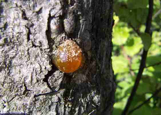
Propolis is a resinous substance that bees collect from tree and plant resins (it looks like red pollen baskets on their hind legs). The bees use this sweet mixture to protect their hive and keep it germ free.
Bees even use the powerful antibacterial benefits of propolis to embalm any hive intruders that the little bees can’t remove on their own. If, for example, a mouse snuck into the hive, the bees would mummify it with propolis to ensure that it doesn't spread its germs and stink up their home!
Propolis through history
Humans have used propolis for centuries dating back to the ancient Greeks and Egyptians. The Greeks used it to treat wounds and abscesses, the Assyrians used it to fight tumors and the Incans drank it to help reduce fever. The legendary Aristotle was into it and is credited by many for coining the term ‘propolis’.
Propolis has been used by many different cultures for diverse reasons throughout history and today many people use propolis to fight colds and sore throats, improve their oral health, prevent inflammation and maintain good health. Ointment containing propolis is also considered by some to be effective in treating herpes or shingles.
So, how can it help?
Immune System
You know that feeling of a scratchy sore throat? The kind that makes speaking painful and enjoying food a no-go? So do I. But there is hope!
Many use propolis to relieve sore throats and to boost the immune system. For that annoying sore throat, the anti-inflammatory and antibacterial activities of propolis can play an important role in soothing the affected area.
When it comes to cold and flu, propolis can help support the immune system.
These are the powerful effects that motivated me to start Beekeeper's Naturals and create a spray that's perfect for those who want to get healthy on the go!
Oral health
Ever lean in for the kiss and get the cheek? You can better your odds of getting some lip service with the help of bee propolis!
Unlike gum, which acts as a cover up, the antibacterial and antifungal properties of propolis get to the root of the problem and fight halitosis (bad breath causing bacteria). In fact, propolis has also been shown to be an effective secondary treatment for gingivitis and plaque.
Many of the large oral care companies have taken note of the effects of propolis on oral health and even launched toothpastes with propolis as a key ingredient!
Antioxidants
Antioxidants are on a lot of people’s minds these days. There is some evidence that active free radical and oxidative stress can contribute to the development of chronic and degenerative diseases.
Propolis contains bioflavonoids and polyphenols, compounds with antioxidant properties that scavenge those pesky free radicals and have an anti-inflammatory affect on the entire body.
Propolis is one of the strongest natural antioxidants out there scoring very high on the Oxygen Radical Absorbance Capacity (ORAC) index, which is often used to compare the antioxidant activity of different foods.
To get your antioxidants from propolis, any form will work from our propolis spray to dried propolis capsules...just don't go eating propolis toothpaste!
In summary...
Propolis is deservedly making its way into the medicine cabinet of international consumers. Check out this natural health helper brought to us by the bees to keep you feeling your best all year round!
Honeybee microbiome is stabilized in the presence of propolis
Honeybees have developed many unique mechanisms to help ensure the proper maintenance of homeostasis within the hive. One method includes the collection of chemically complex plant resins combined with wax to form propolis, which is deposited throughout the hive. Propolis is believed to play a significant role in reducing disease load in the colony due to its antimicrobial and antiseptic properties. However, little is known about how propolis may interact with bee-associated microbial symbionts, and if propolis alters microbial community structure. In this study, we found that propolis appears to maintain a stable microbial community composition and reduce the overall taxonomic diversity of the honeybee microbiome. Several key members of the gut microbiota were significantly altered in the absence of propolis, suggesting that it may play an important role in maintaining favourable abundance and composition of gut symbionts. Overall, these findings suggest that propolis may help to maintain honeybee colony microbial health by limiting changes to the overall microbial community.
1. Introduction
Honeybees (Apis mellifera) as eusocial, cavity-nesting organisms have evolved many mechanisms to maintain a homeostatic nest environment. For proper development of larvae and pupae, temperature and humidity need to remain relatively constant. In addition, the nest architecture must support comb attachment, be waterproof and restrain detritus. Finally, it is critical that microbial growth be controlled in such an environment. Honeybees collect and deposit plant resins on the hive walls to help maintain optimal nest conditions and use these resins to restrict nest entrances to reduce predation and parasitism [1]. As resin is brought into the hive, it is mixed with varying amounts of bee-produced wax and is then termed ‘propolis’ [1,2].
Propolis not only has an architectural purpose but it also functions as a type of social immune defence—a colony-level defense mechanism against pathogens and parasites that arises due to the collective behavior of individuals [3–6]. Resins are collected as a type of social medication with foragers increasing resin collection when the colony is pathogen challenged [7–9]. In addition, the presence of a propolis envelope in pathogen-challenged colonies appears to increase the antimicrobial activity of the glandular secretions that workers feed to developing larvae [10]. Furthermore, bees from healthy colonies with more propolis in the nest interior have decreased investment in their immune response, which may lead to increased lifespan [11]. As reviewed in Simone-Finstrom et al. [5], propolis has both direct effects against pathogens and more subtle effects on individual bees that may translate to reduced disease at the colony and individual levels. However, the specific mechanisms explaining these effects are yet to be elucidated.
Plant exudates incorporated into propolis are rich in secondary metabolites that are not involved in primary plant biochemical processes such as growth and development, but are important for mediating interactions with other organisms (e.g. insects, microbes) [12]. The most well-characterized plant secondary metabolites are essential oils (EOs), which are complex mixtures of volatile aromatic compounds. Natural EO mixtures have a broad range of antiseptic and antimicrobial activities due to the fact that the differing components often exhibit multiple modes of action [12]. EOs, thus, exert pressure on animal microbiota to tolerate, use, or detoxify secondary plant compounds that are encountered in the environment [13,14]. EOs have been experimentally shown to increase host weight gain and improve resistance to infection in some animals via microbiota alterations that impact community structure and function [15].
The honeybee gut and hive environments are colonized by distinct microbial communities that impact individual and colony-level health. Therefore, the collective honeybee gut microbiota and hive microbiota can be considered an extension of the colony phenotype [16–18]. The gut harbours a core community that includes ubiquitous animal gut bacteria Lactobacillus and Bifidobacterium) as well as specialized clades that are shared with other corbiculate bees Snodgrasella and Gilliamella) [17–19]. Gut bacteria are present at low levels in hive materials such as food stores and comb, but core hive bacteria Bombella apis (formerly Parasacharribacter apium [20]) and L. kunkeii) can survive the extreme conditions of the hive and therefore are present at significantly higher concentrations [21]. Both gut and hive bacteria are adapted to survive bee social immune defenses such as glucose oxidase production and innate immune functions such as antimicrobial peptides [17,22]. Even though honeybees do not consume propolis, we hypothesized that the honeybee microbiota has co-evolved to thrive in the presence of propolis. In this way, we postulate that the role of propolis as a social immune defense may extend to influencing microbial homeostasis in the hive and collective gut of the colony.
Since it has previously been documented that a propolis-enriched environment influences total bacterial loads of honeybee colonies [6], the goal of this study was to explore how propolis may specifically influence microbial community structure in honeybees. As our knowledge of the importance of the honeybee microbiota to bee health has increased rapidly over the last several years, investigations regarding how the hive environment influences honeybee microbiota have increased significance.
2. Methods
(a) Environmental parameters and study design
Experimental design was described previously by Borba et al. [23]. Briefly, 12 colonies were provided with commercially available propolis traps (Mann Lake Ltd, MN, USA) stapled to the four inner walls of each bee box to encourage the bees to construct a propolis envelope within the nest. This treatment resulted in propolis-rich colonies. Twelve additional colonies served as controls; no propolis trap was provided and the bees deposited propolis in the cracks and crevices within the box and were left with smooth interior walls, and therefore were propolis-poor. Experimental measures quantifying population and brood size, Varroa infestation, Nosema spp., bacterial load and viral titre can be found in Borba et al. 2015 from the 2012 September cohort [23]. There were no differences in colony size, parasite or pathogen load, or colony bacterial load (as assessed by 16S gene expression) in the colonies at the time of sampling for the current study. However, immune-gene expression was different between the two colony treatments [23], consistent with previous work [6].
Newly emerged bees (noted by their location near eclosing adults from pupal cells, and by their fuzzy appearance [24]) were painted using enamel paint markers and collected after six days [25]. The marked 7-day-old bees were stored at −80°C until analysis. Seven-day-old bees were chosen for analysis as differences in total bacterial loads in bees collected from propolis-rich or propolis-poor colonies were previously observed at this time point [6]. This is also after the age at which the characteristic gut bacterial communities are established [26].
(b) Sample processing and DNA extraction
An average of five to six whole bees from six propolis-rich and six propolis-poor colonies (total N = 62 samples) were processed individually. DNA extraction was carried out following established methods [27,28]. Briefly, whole bees were flash-frozen in liquid nitrogen and ground using sterilized pestle into a fine powder. Following grinding, Tris–EDTA and lysozyme (at 20 mg ml−1) were added and the sample was incubated at 37°C for 30 min. After incubation, proteinase K (at 50 mg ml−1) was added and the sample incubated at 50°C overnight. Phenol–chloroform extraction was performed twice before ethanol precipitation and DNAs were re-suspended in Tris–EDTA and cleaned using a column-based genomic clean-up kit (Zymo) according to manufacture instructions. DNAs were quantified using a NanoDrop2000 instrument (Thermo Scientific Inc., Grand Island, NY, USA). PCR was performed using barcoded Illumina primers following the Earth Microbiome protocols [22,23], with HF Phusion polymerase mix (New England BioLabs, Ipswich, MA, USA) and 3% dimethylsulfoxide (DMSO). Amplifications were performed in triplicate and pooled before normalization based on PicoGreen quantification.
(c) 16S rRNA gene amplicons community analysis and data filtering
PCR and sequencing were performed using a modified version of the protocol presented in Caporaso et al., adapted for the Illumina MiSeq 300 bp paired-end sequencing. The V4 region of the 16S rRNA gene was amplified with region-specific primers that included the Illumina flowcell adapter sequences [27]. Samples were multiplexed and then sequenced on a single flowcell. The software Quantitative Insights Into Microbial Ecology (QIIME 2), an open-source microbiome data science platform, was used for data preprocessing and sequence analysis [29]. Raw sequence data files were de-multiplexed and low-quality reads were removed using the default parameters for QIIME2 for paired-end data. Chimeric sequences were corrected using the DADA2 plugin [30]. Alpha and beta diversity analyses were done using the q2-diversity plugin and to generate the principal coordinates analysis with a minimum sampling depth of 15 000 reads and concordance of features shared by at least two samples. Taxonomic analysis was done with the q2-feature-classifier plugin to map sequence data to taxonomic features. Classifiers were trained directly from the sample data with a 99% similarity to the Silvia 16S sequence database [31]. Representative features were then subject to BLASTn query using the NCBI database to assign specific isolates. QIIME2's diversity plugin was used to perform permutational multivariate ANOVA (PERMANOVA) and a test for homogeneity of multivariate dispersions (PERMDISP) to determine statistical differences in group clustering and dispersion. Identification of differentially abundant features across samples was done using the statistical framework Analysis of Composition of Microbes (ANCOM) and Gneiss [32,33].
3. Results
(a) Bacterial sequences and classification
We obtained 3 103 363 reads sequenced from the 16S rRNA V4 region from 62 samples. Quality filtering reduced the total number of samples to 44 (23 propolis-rich, 21 propolis-poor), with a sequences per sample range from 15 208 to 165 115 (median = 39 448). Sequences clustered in a total of 415 unique taxonomic features. Sampling depth was sufficient to capture similar levels of feature diversity for both treatment groups (electronic supplementary material, figure S1).
(b) Taxa identification
Sequenced reads were resolved down to the family or genus level to maintain confidence in taxonomic designation. The main taxa present across both groups are presented in figure 1a and include taxa such as Snodgrassella, Bartonella, Lactobacillius, Bifidobacterium, Frischella and Gilliamella (figure 1a). These taxa are considered part of the dominant bacteria of the honeybee gut [26,28,34–36]. Figure 1. (a) Taxonomic composition of the microbial communities of bees from propolis-rich and propolis-poor colonies; (b) comparison of the number of times each taxa was observed by a given read (feature abundance) of four significantly differentiated taxa by ANCOM analysis.
(c) Microbial community differences
PERMANOVA and PERMDISP identified significant differences in microbial communities between bees from propolis-rich and propolis-poor colonies using two different dissimilarity measures (PERMANOVA: Bray–Curtis: p = 0.027, r2 = 0.06, weighted UniFrac: p = 0.002, r2 = 0.15; PERMDISP: Bray–Curtis: p = 0.045, weighted UniFrac: p = 0.004). All alpha diversity and unweighted UniFrac distances between the two sample groups were not significant. Principal coordinate analysis (PCoA) plots for Bray–Curtis and weighted UniFrac shows microbial community diversity with significant clustering of the propolis-rich group compared to propolis-poor (figure 2a,b). Figure 2. (a) Principal coordinates analysis (PCoA) using Bray–Curtis and (b) weighted UniFrac measures of dissimilarity showing greater levels of similarity (clustering) among propolis-rich samples as compared to samples from propolis-poor colonies along with the 95% confidence interval indicated by the respective circles. Significance determined through PERMANOVA: Bray–Curtis: p = 0.027, weighted UniFrac: p = 0.002.
(d) Taxa differences
Identification of taxa that were differentiated between bees reared in the propolis-rich versus propolis-poor environments was done using the ANCOM and Gneiss methods. Two taxa (Bartonella and Lactobacillus) were identified as significantly differentiated by both methods. Bartonella and Lactobacillus were both more abundant in the propolis-poor group as compared to the propolis-rich group. ANCOM analysis found a total of eight taxa that were significantly differentiated between the two treatments (Bartonella, Lactobacillus, Bifidobacterium, Enterobacteriaceae, Bombella, Corynebacteriales, Methylobacterium and Dietzia) (figure 1b). However Bombella, Corynebacteriales, Dietzia and Methylobacterium had extremely low abundance and differences could be artefacts of sampling (electronic supplementary material, figure S2). Bartonella and Lactobacillus were more abundant in bees from propolis-poor colonies, while Bifidobacterium and Enterobacteriaceae were more abundant in bees from propolis-rich colonies (figure 1b). The Gneiss method identified three significantly differentiated taxa (Bartonella, Lactobacillus, Snodgrassella (electronic supplementary material, figure S3)). Bartonella and Lactobacillus were more abundant in the propolis-poor treatment (as also indicated by ANCOM analysis), while Snodgrassella was enriched in the propolis-rich treatment.
4. Discussion
We examined the microbial community composition of honeybees in propolis-rich and propolis-poor environments. As bees collected from propolis enriched environments have previously been shown to harbour fewer bacteria (based on 16S rRNA abundance [6] but see also [23]), our aim here was to determine the effect of propolis on specific taxa of the honeybee microbiota. The honeybee microbiota from propolis-rich colonies were more similar to each other in taxonomic composition, compared to propolis-poor colonies. We found that the honeybee microbiota was more consistent between bees collected from propolis-rich colonies, while those from propolis-poor colonies exhibited greater taxonomic diversity. Several bacterial groups were also found to have different relative abundances with respect to the amount of propolis in the colonies. The results presented herein suggest that propolis may support regulation of colony microbiota by maintaining a stable or homeostatic microbial community.
Bray–Crutis and weighted UniFrac measures of dissimilarity found significant differences in the overall taxonomic microbial diversity between bees from propolis-rich and propolis-poor colonies. Significance in the PERMANOVA tests demonstrates that there were overall localized differences between the two groups across the two diversity measures. PERMDISP provides additional evidence that the two groups were also significantly different in terms of the variance distribution that exists within propolis-rich and propolis-poor colonies. The latter of the two tests provides subsequent evidence that the microbial population variation is limited in the propolis-rich colonies and thus more stable across colonies as compared to the propolis-poor colonies. Given that honeybee-associated microbes have likely co-evolved with their hosts in the presence of propolis, as propolis use is ubiquitous in feral colonies nesting in tree cavities [1], some microbial symbionts may be more or less sensitive to its chemical properties. While this has yet to be explored, these results raise the question of whether specific ratios of particular taxa are particularly important for bee health or if the stability of representation of specific taxa is key to preventing dysbiosis [19]. While it is likely a combination of the two, variation in community composition and diversity can dramatically affect the overall health of the host [37]. Four of the eight significantly differentiated taxa we identified in this study are considered to be core members of the honeybee gut microbiome (Lactobacillius, Bifidobacterium, Bartonella and Snodgrassella) [34]. Lactobacillus and Bifidobacteriaceae both belong to taxonomic groups that are implicated in carbohydrate transport and polysaccharide processing [38]. Lactobacillus were primarily comprised of the ‘Firm-5′ phylotype, and understanding how its differential abundance with respect to propolis or other environmental conditions could impact colony health is important for future work. Bifidobacteriaceae were more abundant in the propolis group and have been previously shown to be important to maintaining colony health [17,26,35,37,39]. Bartonella is a gut symbiont [40] that has been shown to differ seasonally [41] and in honey bees exposed to various types of landscapes [34], raising the question of its functional roles and how propolis may interact with it.
Other significant taxa identified in the study, such as Enterobacteriaceae, are facultative anaerobes that are suggested to be involved in metabolic processes such as including sugar and nitrogen processing [41]. Therefore, alterations in the abundances of this family of microbes may have an impact on host metabolism. Snodgrassella was the most abundant taxa identified in our study. Snodgrassella is a common honeybee symbiont which modulates the gut environment by consuming O 2 to maintain anaerobic conditions in the gut lumen [19,42]. The significant abundance of this bacterium in the bee gut suggests that it may play a significant role in maintaining the overall homeostasis of the gut microflora. Although we were unable to detect strain-level differences within Snodgrassella, it may be interesting to explore how the specific strains regulate gut community dynamics and host physiology. Further study is also necessary to better characterize the specific effects of microbiota shifts and how they are related to functional changes in host physiology and health (see electronic supplemental material, PICRUSt analysis), particularly in this context of strain diversity.
5. Conclusion
In total, our results demonstrate that honeybees in a propolis-rich environment differ in their relative abundances of core microbial community members. These findings provide interesting and novel insight into how a feature of the nest environment can influence the community structure of co-evolved bee-associated microbes, and suggest an additional mechanism by which propolis may contribute to overall colony health. Future work should address if disruption of gut microbiome homeostasis influences the establishment of pathogens or growth of opportunistic species and if this is mediated by differential responses to the antimicrobial activity of propolis or indirectly through the bees' physiological responses to propolis in the hive environment. Additionally, we have identified candidate taxa for future functional investigations that may help to further understand complex microbial dynamics involved with regulating the health of honeybee colonies.
Data accessibility
The data underlying this study are available from Dryad: [43].
Authors' contributions
The study was designed by R.S.B., M.S.F. and M.S. Sample collection and preparation were done by R.S.B. and M.S. Sequencing data were analysed by P.S., and results were interpreted by P.S., M.S.F. and V.R. P.S., M.S.F. and V.R. wrote the manuscript and all authors provided edits and approved the manuscript. All authors agree to be held accountable for the context herein.
Competing interests
The authors have no competing interests. Any mention of trade names or commercial products in does not imply a recommendation or endorsement by the USDA. USDA is an equal opportunity provider and employer.
Funding
Funding for this work was provided to M.S. by NSFIOS-1256992, to M.S. and M.S.F. by USDA-NIFA2018-67013-27532, and to a USDA postdoctoral fellowship to M.S.F.
Acknowledgements We thank Irene Newton for initial method consultations and the thoughtful anonymous reviewers for their comments that significantly improved the final version of this manuscript.
Footnotes
†Authors contributed equally. Electronic supplementary material is available online at